Technology to preserve brain function when removing tumors
Executive Briefing
Synthesized and verified with gemini-2.0-flash-thinking-exp
“# Technology to preserve brain function when removing tumors Technology supports mapping, finding t”
Key Takeaways
- 1.# Technology to preserve brain function when removing tumors Technology supports mapping, finding the way to the tumor, warning of risks and magnifying the surgical field, helping to safely dissect the tumor and protect the patient's brain function.
- 2.Le Van Ngan, Department of Neurosurgery - Spine, Tam Anh General Hospital, Hanoi, four layers include 3 Tesla MRI combined with DTI to map the tumor and nerve fiber bundles; Neuro-Navigation determines tumor location, instruments and access; Intraoperative electrophysiological neuromonitoring system (IONM) monitors functional signals and warns of risks; Zeiss Kinevo 900 robotic microsurgery magnifies the surgical field, supporting the separation of tumor tissue from blood vessels, nerves and healthy brain tissue.
- 3.The four systems are applied in series, with data from the previous layer supporting decisions at the next layer.
Technology supports mapping, finding the way to the tumor, warning of risks and magnifying the surgical field, helping to safely dissect the tumor and protect the patient's brain function.
According to MSc. Le Van Ngan, Department of Neurosurgery - Spine, Tam Anh General Hospital, Hanoi, four layers include 3 Tesla MRI combined with DTI to map the tumor and nerve fiber bundles; Neuro-Navigation determines tumor location, instruments and access; Intraoperative electrophysiological neuromonitoring system (IONM) monitors functional signals and warns of risks; Zeiss Kinevo 900 robotic microsurgery magnifies the surgical field, supporting the separation of tumor tissue from blood vessels, nerves and healthy brain tissue.
The four systems are applied in series, with data from the previous layer supporting decisions at the next layer. The technology chain is especially useful for patients with brain metastasis, when doctors need to remove the tumor, relieve pressure but still preserve brain function so the patient can continue cancer treatment.
"The ultimate benefit of the four layers of technology therefore lies not only in the amount of tumor removed, but also in the patient's ability to maintain movement, language, cognition and level of independence after surgery," said Dr. Ngan.
Doctor Ngan cited the case of Mr. Hau, 67 years old, treated at Tam Anh General Hospital in Hanoi. The patient has late-stage lung cancer, has a metastatic tumor in the right frontal lobe, measuring 22 x 27 x 31 mm, compressing the brain, causing the left side of the body to gradually weaken.
The challenge of the surgery is to remove the tumor but avoid motor pathways in the adjacent brain area. Therefore, the team applied four layers of technology, in turn answering the questions: where is the tumor located, which way should be approached and whether the current operation will affect function or not.
The first layer is used before the doctor opens the skull. 3 Tesla MRI provides images of the tumor and surrounding structures. Diffusion Tensor Imaging (DTI) technique analyzes the direction of diffusion of water molecules along the axon, thereby building a three-dimensional model of some nerve conduction bundles.
The map shows the relationship between the tumor and nerve fiber bundles responsible for movement, sensation, vision or language. For Mr. Hau, the data helps the team identify tumor areas near motor pathways and plan approaches to avoid structures that need to be preserved.
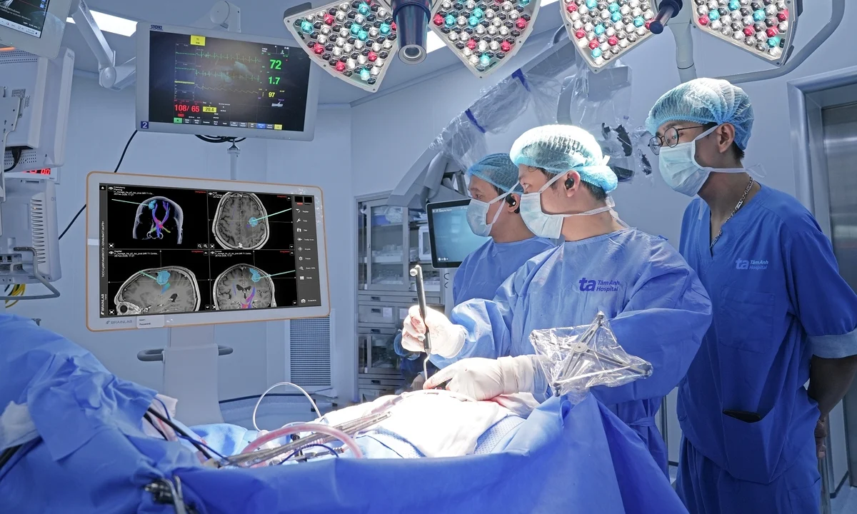
The second class brought the map into the operating room. Neuro-Navigation uses cameras to monitor reflective points mounted on the patient's head, positioning rods and surgical instruments. The software compares real anatomical landmarks with MRI data, displaying the instrument tip position on a three-dimensional brain model.
When the doctor moves the probe on the surface of the head or in the operating field, the screen shows the corresponding position, distance to the tumor and built-up fiber bundles. The team can simulate many approach angles, thereby choosing the appropriate skull opening location and brain access route.
However, positioning does not guarantee that the data always matches perfectly. After opening the dura mater, aspirating cerebrospinal fluid or removing part of the tumor, the brain may shift compared to the preoperative scan, a phenomenon called brain shift. Therefore, the team needs additional functional signals that arise during the surgery to test the safety limit.
The third layer is the intraoperative neuroelectrophysiological monitoring (IONM) system. The electrodes are placed at appropriate locations on the body, and can combine brain stimulation. The system continuously records the response of the transmission paths; The specialist monitors changes in amplitude and conduction time on the screen.
In Mr. Hau's case, IONM assisted the team in choosing the brain entry point to avoid the motor area and monitoring the signal throughout the tumor removal process. On the screen, the green line represents the baseline EMG amplitude, the white line is updated during surgery. The hospital said that the signal after tumor removal did not decrease compared to baseline, showing that motor transmission pathways were preserved; The surgeon does not have to adjust the operation because of the IONM warning.
If the signal decreases, the team must check the cause, from the position of the tool, the level of tissue stretch, blood perfusion to anesthesia factors, then adjust or pause the operation when necessary. IONM therefore does not create additional anatomical images but acts as a functional warning channel, supplementing MRI maps and navigation systems.
The last class is located at the operating theater. The Zeiss Kinevo 900 robotic microsurgery magnifies the surgical area, helping doctors observe blood vessels, nerves, tumor tissue and brain tissue through narrow access, while also changing and saving viewing angles. In Mr. Hau's case, glasses assisted the doctor in dissecting under direct observation.
After surgery, Mr. Hau had no new neurological dysfunction, his left side weakness improved and he could take care of himself. He was discharged after 7 days and continued treatment with targeted drugs. Three months after surgery, the patient's health is stable.
Before Mr. Hau's case, Tam Anh General Hospital in Hanoi performed 50 brain tumor surgeries in 2025, all using MRI DTI, Neuro-Navigation and microsurgical glasses. Of these, 15 cases had tumors located in important functional areas that were supplemented with IONM, which means using all four layers of technology. More than 90% of patients no longer see tumors on MRI after surgery. About 5-10% have neurological dysfunction, mainly temporary.
Dr. Ngan assessed that the four layers of technology did not completely eliminate risks or decided to change surgeons. The value of the system lies in continuously providing information about anatomy, instrument position and nerve function, helping doctors adjust each operation according to actual developments. The goal of brain surgery is therefore not only to remove the maximum tumor but also to preserve the patient's mobility, communication, cognition and self-care abilities.
Dragon Fruit